


The valgus deformation of the legs is most often congenital. However, in some cases, paralysis, traumatic lesions - may appear in mature life. The main symptoms of pathology are pain in the legs and lower leg muscles, visible violations of the shape of the legs, and changes in walking. Diagnosis of the disease clinical examination, X -try, electromiography, etc. Handling using treatment. The treatment includes conservative and surgical methods. However, proper efficiency is observed only in the reconstruction operations.
What is this disease?
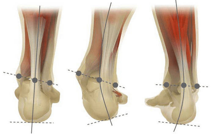
Valgus deformation is the curvature of the foot characterized by rebounds of the longitudinal arc. Usually the inner edge of the foot is lowered ("drops") and the heel turns out.
At the foot of a person, due to its location, he takes the pressure of the entire mass of the human body. Therefore, it has a special anatomical structure that allows depreciation, balancing and stabilizing movements. However, the correct stop form is an important element of the tasks.
Nowadays, traumatology and orthopedic are the most important problems with the foot. The meeting estimates that 30-58%, where the cases make a congenital disorders on 2/3.
Pathology is largely socially significant because it covers all age groups of the population and promotes the drive of the spinal column, the early development and arthrosis of the lower limbs.
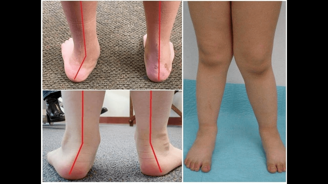
If you pick up your leg (if you look at them from the back), an ankle level develops x-like deformation: the ankle is in contact, while the heel is 5-6 centimeters apart.
The pathology is most often congenital and is diagnosed in children in the hospital (or immediately after the start of the walk). A similar condition is adapted for 5 years after (in the absence of proper treatment) a child develops flat legs.
Why does it arise?
It is believed that the main reason for the appearance of the valgus deformation of the foot is the inadequate function of the back tibial muscle or the weakness of the league.
Nowadays, other factors of the development of pathology can be distinguished:
- Congenital disorders with the improper location of the leg bones or shortening of the tendons (vertical ram bone, short pile of tendons);
- Posture disorders If the deformation of the legs compensates for the curvature of the spinal column;
- Traumatic lesions (fractures of the leg bones, lower legs, hips or knees, league and tendons);
- Paralysis (immobilization) due to nervous system damage due to damage to violation of cerebrospinal melting, etc. ;
- Cramping (constant contraction) of the lower leg muscles;
- Concomitant diseases: the pathology of the bone system with vitamin D (Rickkets), diabetes mellitus, osteoporosis (reduction of bone density), impaired thyroid glands and parathyroid glands, etc. ;
- Increased body weight, including the rapid increase in menopause or during pregnancy.
Pathology is also facilitated by incorrectly selected shoes or excessive correction of the club foot in childhood.
The degree and stage of the disease
The severity of pathology (the power of manifestation) is divided by a degree:
- Light, 1, 5-2 centimeters of vault height and heel inclination to 15 degrees;
- The average when the arc is hit to the 1 centimeter and the angle decreases to 10 degrees;
- It is difficult at the height of the vault up to 0, 5 cm and the angle of the heel is 5 degrees.
Depending on the involvement of certain structures, the following stages of curvature can be distinguished:
- There are no bone deformations, pain should be determined on the inner surface of the ankle (in the area of the back tibial muscle);
- The curvature is light, the corner is slightly rejected;
- The leg is distributed and the deformation is fixed (not properly corrected);
- The run is observed not only in the foot but also in the ankle joint.
Symptoms
In the first phase, patients with periodic pain interfere with long -term walks or long vertical loads (standing or at the leg). As a general rule, the pain syndrome increases when it is not properly selected. The next stage of the disease is associated with the occurrence of the leg curvature: patients in standing position do not rely on the outer edge of the foot, but throughout the area. Notice the slight change in walking.
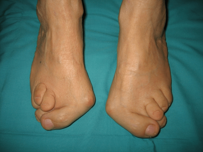
In the third stage, we determine the protrusion of the melted bone (noticeably lower than the ankle on the inner surface of the ankle) and a strong diversity of the heel (the patient is based on the inner edge of the heel bone). The advanced valgus deformation of the legs is characterized by the pronounced curvature of both the leg and the ankle joint. Patients complain of severe pain in the lower leg muscles and significant walking violations: the knee rubs together while the right and left leg are located at a certain distance.
Severe leg curvature of the legs is often complicated by the spinal column (skoliosis with different positions of shoulders and pelvis wings), osteochondrosis (damage to intervertebral disk with hernia) or arthrosis (damage to pre -articular abrasion in the lower leg, knee and hip).
How to diagnose?
Diagnosis of foot curvature consists of:
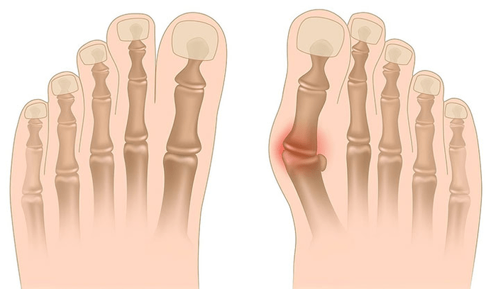
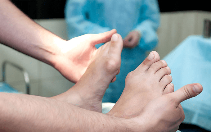
- Clinical control, during which the orthopedist detects a decrease in the arches of the foot, the difference in heel and ram bones, the visible "disappearance" of the outer ankle and the protrusion of the inner ankles.
- X -Ray - an affordable and informative method to determine the change in bone inclination and the linear parameters of their relationships. These indicators are required to make a definitive diagnosis and clarify the degree of deformation.
- Method of registration steps aimed at determining the exact functional state of the limb. The method consists of registering the time of the leg parts of the foot. The study also examines phases of foot rolling, which reflect the balance of the lower limb muscles.
- Dynamic electromiography that records the electrical activity of the muscles tested and its addiction from the step phase.
- Photopymography with digital processing that allows you to obtain all standard indicators and the type/degree of curvature with high accuracy.
Further neurologist consultation (with deformations due to cramps or paralysis), endocrinologist (thyroid/parathyroid diabetes or disorders) and gynecologist (when the threat) may be required. If the leg curvature is against the background of osteoporosis, densitometry is required - examination of bone density.
Treatment
The main methods of treating the valgus curvature of the legs are conservative and operational distinctive. Do not destroy sore joints with ointments and injections!
Conservative approach
This type of help aims to get rid of the symptoms of the disease, but does not eliminate the cause of the pathology.
The technique includes:
- the use of orthopedic insoles to support I plus bone, foot arc and eliminate valley lesions of the middle and back of the foot;
- Taping - Fixing the leg and ankle with special adhesive tapes with proper elasticity. The tape is worn around 3-5 days and replaced;
- Sewing orthopedic shoes according to individual standards;
- The use of orthosis and other fasteners on the leg and ankle.
Conservative methods include a complex of physiotherapeutic procedures (ozokerite, paraffin applications, electrophoresis, magnetic effects), massage and physiotherapy, developed for a particular clinical case. Be careful! Nowadays, most experts prefer surgical treatment methods, as conservative therapy is ineffective (statistics say 60% of cases are useless).
Surgical intervention
The amount and type of operation depends on the direct stage of the disease. Thus, the first degree of Valgus deformation is treated with synovectomy (removal of tendon to correct overall tension) or the osteotomy (autopsy) of the heel to return to anatomically correct position. In the second phase of the disease, transplantation of the bends of the fingers is used. Such an intervention is usually performed by the autopsy of the heel or the ram-Lobed arthrodesis (surgical immobilization of the joint between RAM and Scaphoid Bones).
The III. Grade curvature requires arthrodesis of several joints at the same time: an extra-free, five-fold cubic meter and RAM. Such three lower immobilizations are often complemented by autopsy of the corner bone. The pathology IV. The reconstruction operations are needed not only on the leg but also on the ankle. In this case, the instability of the league device is adjusted by transplantation (from their own bodies or artificial materials). The amount of operations on the foot itself is the same in the III. Curvature.
Recovery period
Rehabilitation includes walking without support on the foot for 2 months. At the same time, the patient should wear a removable plaster length for 1, 5-3 months. After 1, 5 months after surgery, active movements are recommended on the operated foot. Month 3 is introduced a complex to strengthen physical education. However, patients are prohibited by curly walking and active sports activities. It is worth noting that only six months later the final result of the surgery can be judged.
Preventive measures
Preventing a Stop Deform is included in the following measures:
- Early correction of congenital disorders by improper arrangement of the bones of the legs or shortening of the tendons (vertical melted bone, short heel tendon);
- Repair of posture disorders (skoliosis, etc. );
- Timely treatment of traumatic lesions (fractures of the legs, lower leg, thigh or knee joint, league and tendons cracks);
- Correct rehabilitation (immobilization) is due to injury to the injury of nervous system roots due to damage to encephalitis, polyomyelitis, polyomyelitis, stroke, cerebrospinal roots, etc. ;
- Alleviation (constant reduction) of the lower leg muscles;
- Concomitant disease therapy: bone pathologies with vitamin D (rickets), diabetes mellitus, osteoporosis (reduction of bone density), thyroid gland and parathyroid glands, etc. Impaired function;
- Normalization of body weight (especially in the case of postmenopause or rapid weight gain due to pregnancy);
- Selection of orthopedic shoes or using superprins;
- The moderate correction of Clubfoot without "hyper-correction" leads to secondary valgus emptying of the legs.
Preventing the progression of the disease is the use of conservative methods and early reconstruction operations. In this case, physical activity is limited to prevent ankle joints and curvature. Keep in mind that timely treatment of the valgus deformation of the legs not only improves patients' quality of life, but also prevents osteochondrosis and arthrosis of the knee or hip joints!